Flow Cytometry (FCM) is a technique for rapid quantitative analysis and sorting of cells or other biological particles (such as microspheres, bacteria, small model organisms, etc.) arranged in a single row in a liquid stream. As a technology platform for the detection of flow cytometry, modern flow cytometry was produced in the 1960s and 1970s. After nearly 40 years of development and improvement, today's flow cytometry is very mature and widely used in all aspects from basic research to clinical practice, covering cell biology, immunology, hematology, oncology. The fields of pharmacology, genetics, and clinical testing play an important role in various disciplines.
Overview
A technique for rapidly determining the biological properties of individual cells or organelles in a flow system and classifying specific cells or organelles from the population. It is characterized by rapid determination of Coulter resistance, fluorescence, light scattering and light absorption to quantify many important parameters such as cellular DNA content, cell volume, protein content, enzyme activity, cell membrane receptors and surface antigens. Cells of different nature are separated according to these parameters to obtain a pure cell population for biological and medical research. The highest sorting speed has reached 30,000 cells per second.
Modern flow cytometry combines fluid mechanics technology, laser technology, electronic physics technology, photoelectric measurement technology, computer technology, fluorescent chemical technology and monoclonal antibody technology, which is the crystallization of multi-disciplinary and multi-disciplinary technological progress. With the rapid development of modern science and technology, in order to meet the higher requirements of life science for cell analysis, flow cell technology is still developing rapidly, and has made many breakthroughs in detection technology, sorting technology and high-throughput analysis. .
Brief history
In 1934, A. Mordawan first reported an automatic cell counting method for passing suspended red blood cells through a glass capillary placed on a microscope stage. In 1956, WH Kurt introduced a device for performing cell counting and measuring cell volume by using a change in electrical resistance (called Coulter resistance) between two inter-cell pores (75-100 μm) in a conductive solution. . In 1965, LA Kamenzki made a multi-parameter flow cytometer that measures cell size and nucleic acid content. In the same year, MJ Furl Weller made a cell sorter. In 1969, Van Dilla et al. used an argon ion laser and a layered shell flow technique to establish a flow cytometer with liquid flow, illumination optical axis and detector axis orthogonal to each other.
Later, the improved cell sorting meter by HR Hewlett enables the cells in the flowing liquid to be ejected into the air for measurement. However, in each of the above systems, the laser beam used and the limiting diaphragm provided in the direction in which the fluorescence is collected are larger than the cells in the flow, and thus cannot provide information on the morphology of the cell, so it is called zero resolution. system. Subsequently, LL Huilis and SF Patten developed a low-resolution slit scanning technique that measures the nuclear fluorescence, cell and nucleus size. In 1969, W. Ged and W. Dietrich described a flow cytometer using a mercury lamp as a light source to excite cells that flow parallel to the optical axis in the flow chamber under epi-illumination.
principle
The sample to be tested (such as cells, chromosomes, sperm or bacteria) is dyed by a fluorescent dye to form a sample suspension, which enters the flow chamber through a sample tube surrounded by the shell liquid under a certain pressure, and is arranged into a single column of cells, which is flowed. The nozzle of the chamber is ejected to become a stream of cells and intersects the incident laser beam. The cells are excited to produce fluorescence, which is collected by an optical system placed at 90° to the incident laser beam and cell fluid. A blocking filter in the optical system is used to block the excitation light; a dichroic beam splitter and other blocking filters are used to select the fluorescence wavelength. The fluorescence detector is a photomultiplier tube. The scattered light detector is a photodiode that collects forward scattered light. Small angle forward scatter is related to cell size.
The entire instrument processes the fluorescent pulse signal and the light scattering signal with a multi-channel pulse height analyzer. The results of the measurements are represented by a single parameter histogram, a two parameter scatter plot, a three dimensional view, and a contour (contour) plot.
The principle of sorting cells is that high-frequency oscillation is generated by the ultrasonic oscillator to vibrate the flow chamber, and the flow of the cells ejected from the nozzle is broken into a series of uniform small droplets, and some droplets contain cells. Before the cells form droplets, the optical system has determined their signal (representing the nature of the cell) if the measured signal matches the selected cell properties to be sorted, or if it is found to be classified When the selected cells are just forming droplets, the instrument charges the entire stream with a short positive or negative charge. When the droplet leaves the stream, the droplets of the selected cells carry a charge, while the selected cell droplets are uncharged. The droplets with positive or negative electricity are deflected toward the cathode or toward the anode when passing through the high-pressure deflector, thereby achieving the purpose of classifying and collecting cells.
application
Cell biology research
Flow cytometry can be used to determine the percentage of cells in each phase of the cell cycle. The DNA content distribution curve is obtained by measuring the DNA content of each cell in the cell population. For example, in determining the DNA content distribution curve of Hela cells, the first peak is G1/G0 (pre-synthesis/quiescent phase) cells containing 2CDNA content, and the next peak is G2+M of 4C DNA content (late DNA synthesis + mitosis) Period) Cells, from the 2C to 4C region, are S phase (DNA synthesis phase) cells. The percentage of cells in the cell cycle at each cell population can be calculated by plotting or computer fitting.
Multi-parameter analysis can be performed by flow cytometry, which simultaneously measures multiple properties of a cell. Such as scattered light and fluorescence, or a variety of different colors of fluorescence. For example, after the cells are stained with acridine orange, the DNA is green and the RNA is red. By measuring these two kinds of fluorescence, it is possible to simultaneously know the DNA and RNA content in one cell. The measurement results can be expressed in a two-dimensional scattergram or a three-dimensional perspective. In this way, G0 and G1, M and G2 cells can be identified based on DNA and RNA content. Flow cytometry combined with liquid scintillation technology can also determine the time (T, Ts, T+M) and coefficient of variation of cells passing through G1, S and G2+M phases.
In recent years, bromodeoxyuridine which has been infiltrated into cellular DNA can be found by using a monoclonal antibody against bromodeoxyuridine (Brdurd). Combining this fluorescent antibody technology with the determination of cellular DNA content is a very useful technique for studying DNA synthesis and cell cycle. In addition, flow cytometry can also determine the extent and timing of cell population synchronization, identify dead cells and living cells, and use fluorescently labeled ligands to quantify receptors on the cell surface and inside.
Genetic research
The chromosomal DNA content is determined by flow cytometry to obtain a chromosome frequency distribution map (Fig. 2), which is called flow karyotype analysis. A peak appears on the same type of chromosome, and the area of ​​the peak represents the abundance of this type of chromosome. Flow chromosomal karyotyping technology not only can quickly analyze karyotypes, but also sort out different types of chromosomes, and make DNA libraries for each chromosome of humans, which can be used for the research of human genome research, genetic diseases and cancer diagnosis.
Immunology research
In combination with immunofluorescence, flow cytometry can identify and count cells with different surface-specific antigens, such as fluorescein-labeled immunoglobulins to identify T and B lymphocytes (Figure 3), depending on the cell surface antigen, Further distinguishing different T and B lymphocyte subsets, as well as determining the number, density and kinetic parameters of antigens carried by each cell. Cell populations with "+" and a specific antigen without "-" can also be collected by flow cytometry to study their functional properties.
Flow cytometry is an important feature for determining immunodeficiency, such as AIDS (acquired immunodeficiency syndrome), that is, the proportion of T4 and T8 lymphocytes is changed (a large number of T4 cells are reduced), and autoimmune diseases and leukemia and lymphoma are determined. Phenotypes, etc., are very useful. In addition, flow cytometry can also be used to quantitatively analyze fluorescein-labeled lectins bound to cells, determine the relative surface density of cells and the relative density of fluorescein binding sites, and combine cell kinetics to determine each cell binding site. The number, as well as the competition between various exogenous lectins and cell surface binding.
Oncology research
Tumor cells generally contain an abnormal amount of DNA. Aneuploid cells are found in most solid tumors and acute leukemias. Because of the simple preparation method of flow cytometry samples, accurate measurement results, and rapid information on DNA ploidy, it can provide valuable diagnosis. data. If the DNA content is measured, and other parameters (such as different types of medium fibrin, protein content, cell size, nucleoplasmic ratio, etc.) can be further improved, the reliability of the diagnosis can be further improved.
Flow cytometry can evaluate the role of tumor chemotherapy and radiotherapy in both experimental and clinical settings. The task of monitoring tumor therapy based on flow cytometry and cell dynamics data has now been carried out in practice.
In addition, flow cytometry is also widely used in hematology, microbiology, molecular biology and other fields. Flow cytometry is developing toward high sensitivity, high speed, multi-parameter measurements, and acquisition of morphological information.
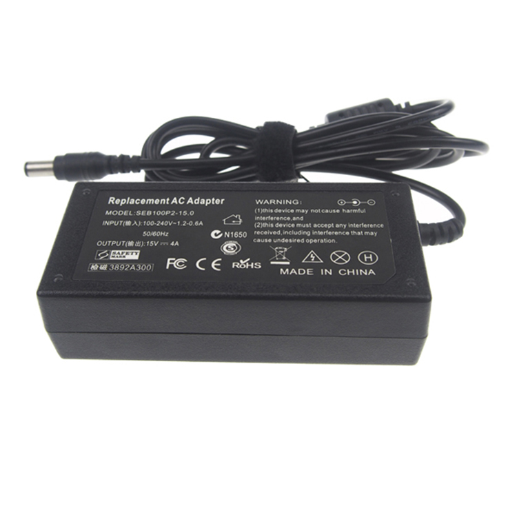
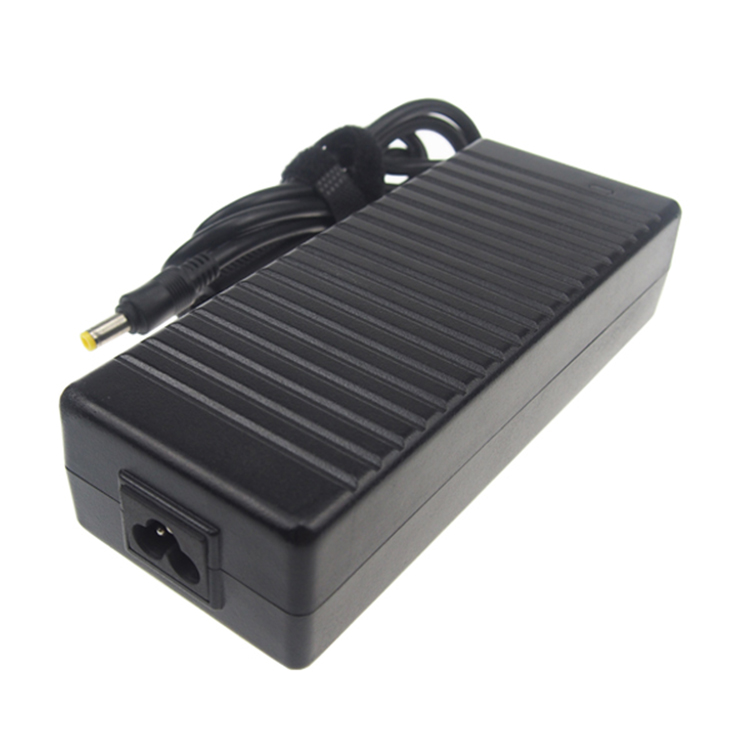
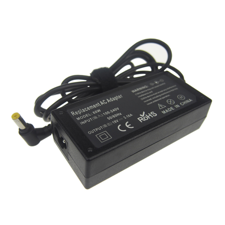
Yidashun offer full replacement NEC power Adapter for laptop with best service at the most competitive prices! All our NEC Laptop Charger are Brand New Replacement Product, works as Genuine parts, 100% OEM Compatible!! Our adapter with smart IC to protect your laptop with over current protection, over load protection, short circuit protection, over heat protection.
If your original NEC laptop adapter is not work anymore, please tell us your laptop model, we will help select the correct OEM replacement models for you. we offer a full 1 year warranty for our adapters.


NEC Laptop Charger,NEC Charger,NEC Adapter,NEC Computer Laptop Charger
Shenzhen Yidashun Technology Co., Ltd. , https://www.ydsadapter.com